 WISHING EVERYONE A HAPPY NEW YEAR! WE'VE RETURNED TO OUR REGULAR OPENING HOURS
What is Osgood Schlatter’s Disease? (OSD)
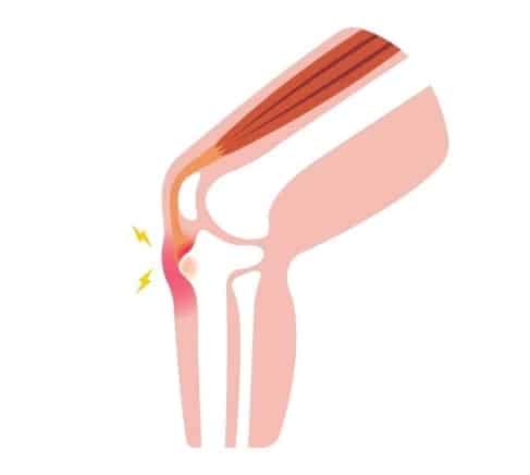
Osgood Schlatter’s Disease (OSD), also known as osteochondrosis, tibial tubercle apophysitis, or traction apophysitis of the tibial tubercle refers to a condition characterized by pain in the tibial tuberosity (front of the knee, just under the knee cap) in adolescents.
As scary as the name may sound, OSD is not a disease, but an overuse condition characterized by ossification of bone along the growth plate, the tibial tuberosity.
Repeated traction and microtrauma (usually from jumping and sprinting) at the tendon insertion cause microvascular tear, inflammation and fracture. This can result in painful swelling at the tibial tuberosity.
Signs and Symptoms
- Gradual onset of pain
- Pain and swelling under knee cap (at the tibial tuberosity)
- Pain gets worse with running, jumping, steps/stairs, and walking uphill
- Limping may be present due to severe pain.
Usually only one knee is affected but sometimes it can affect both knees.
Anatomy:
The tibial tuberosity is an apophysis, meaning that it is a bony prominence that acts as a site for tendon attachment – in this case: the patellar tendon.
The patellar tendon connects the patella (knee cap) and the tibia (shin bone) at the tibial tuberosity.
In adolescents, there is a growth plate at the tibial tuberosity that ossifies (turns into a bone from cartilage) between 7-12 years.
Repeated pulling from the patellar tendon can cause small tears, inflammation, and fractures.
Risk Factors:
- Gender: M > F
- Ages: M (12-14 years), F (10-12 years)
- Sports involving repetitive jumping and sprinting (High intensity/High impact)
- Sudden growth spurt/skeletal growth
- Tight Quadriceps
Treatment:
Non-operative treatment of OSD is based on the same principles that apply all overuse injuries i.e RICE or PEACE and LOVE.
Treatment involves activity modification to reduce bleeding, preventing aggravating the injury, and to allow the tendon and tissues to heal. Examples of activity modification include reducing the frequency, time or intensity of the activities such as training, or of specific movements i.e jumping and sprinting.
Note: activity modification does not always mean complete immobilization. The child should continue normal physical activities within their pain limits. External supports such as knee-braces, strapping and knee compression supports may help during activities to help reduce pain.
Rehabilitation includes gradual progressive overload and gentle stretching to the quadriceps and hamstring.